Regarding recently circulated "Pfizer evidence of graphene oxide" in the injections
Graphene is mentioned in the Pfizer document, but not as part of the injection substance
I was forwarded a FOIAed Pfizer document describing lab studies done by Pfizer Groton CT lab characterizing spike protein as a target antigen for “vaccine” development. The document is 14 pages.
First thing to note - the names of the “contributing scientists” for these experiments as well as those who approved the report are redacted with (b) (6) redaction which we know by now means that the US foreign relations may be damaged if those names are revealed. So, it’s National Security kind of thing. I wonder why, if we are currently allowed to think only 2 thoughts:
-Theory 1: these are regular corporate pharma employees working on a revolutionary new medical product, a “life saving vaccine”; or
-Theory 2: bad Pfizer “captured” FDA, made a bad version of the “life saving vaccine”, made a bunch of false claims and collected big money from the incompetent government.
If either 1 or 2 is correct - why redact the names, and why use (b) (6) as the justification?
Is this a report on the science experiment for a medical product, even if badly made, or a part of the “state-of-the-art weapons system”? If the latter, of course, it makes sense to keep the names secret, as we don’t want the Kingdom of Thailand, for example, to find out the names of those responsible for ending their 785 years old royal lineage, as their 44 yo previously perfectly healthy royal princess collapsed 23 days post Pfizer booster. That would be damaging to the US foreign relations, I think. We also don’t want those people to be targeted/entrapped/abducted/recruited/turned, etc. and put to work by another evil empire in a reverse-Paperclip operation. Our own evil empire must stay competitive.
Updated in March 2024: The royal family in the UK, including King Charles, Kate Middleton and Fergie seem to have been poisoned with the injections, too.
Back to the Pfizer report.
The purpose of this study, conducted in early 2020, was to express SARS-CoV-2 P2 S (“spike protein”) encoded by BNT162b2 (Pfizer’s injection product) for structural biophysical characterization. One type of this characterization was to image the spike protein with an electron microscope and develop a model of the “ideal” spike protein structure folded into its final shape as the antigen target for “vaccine” development.
This study used a variant of HEK cells - human embryonic kidney cells, a “commercial” cell line obtained from aborted fetuses. The cell line was Expi293F™ Cells - human cells derived from the 293F cell line. They are maintained in suspension culture and will grow to high density in a proprietary expression medium. These cells are highly transfectable - they take up a lot of genetic material they are bathed in and grow a lot of target protein quickly, within 48 hrs.
After it is grown, the spike protein is purified, sort of: P2 S with molecular weight of 150 kDa, as well as dissociated S1 and S2 subunits (75 kDa), were used in the structural characterization. The report noted that spontaneous dissociation (breaking off) of the S1 and S2 subunits occurs throughout the course of protein purification.
They did experiments on P2 S binding to human ACE-2 receptors and found that P2 S bound to the human ACE2-PD, and an anti-RBD human neutralizing antibody B38 (from Covid-19 recovered patients) with high affinity.
Next experiment was electron microscopy imaging. This is where graphene oxide is mentioned in the set up of the study (p.7):
Many thanks to Michael Palmer, MD/PhD, for translating this into English for me. Apparently graphene oxide was used to coat the mesh grid holding the spike protein when imaged.
This paragraph contains the technical details of their cryo-electron
microscopy studies for structural characterization of the spike protein.
The purpose is the same as with protein crystallography, but you don't
need to grow crystals first. Instead, you can just dump a liquid sample
containing purified protein onto an EM sample holder (mesh grids), then
deep freeze it in order to minimize thermal motion/fuzziness and
collect a large number of EM images in phase contrast mode. This images
are then all aligned and averaged in the computer in order to extract a
not-quite-atomic 3D image of the ideal molecular particle.This image is then used as a "cast" into which the known protein
sequence is shoved in order to generate a model of the structure with
actual atomic resolution.
So that is the purpose here -- create an experimentally supported
high-resolution 3D model of the spike protein.
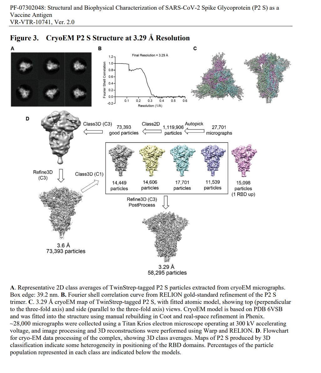
And this is what the CryoEM images and subsequently modeled structure look like:
Note that this is what the “vaccine” supposed to induce your cells to make - this shape and size of the protein. And we know it may or may not do that. Most of the time it makes something else, in fake CMC documentation submitted by Pfizer to the EMA a different size of protein was made, a much larger one - 180-230 kDa, according to what appears to be faked Western blotting technique, and no corresponding model of the structure was produced. So we still don’t know quite what kind of protein is produced, but we know it is damaging to the victims.
Bottom line, nobody knows what is being made in the cells of the unfortunate victims of the mass-scale unrestricted warfare, of which this chemistry is only part. A state-of-the-art part.
Does this prove graphene oxide in the injections?
NO. It just proves Pfizer uses graphene oxide at Groton labs for CryoEM high resolution imaging sample prep for this study.
A recently published paper is describing this imaging technique. Note that Pfizer has close R&D collaboration with U Mich at Ann Arbor.
Cryogenic electron microscopy (cryo-EM) has become a widely used tool for determining protein structure. Despite recent technology advances, sample preparation remains a major bottleneck for several reasons, including protein denaturation at the air/water interface, the presence of preferred orientations, nonuniform ice layers, etc. Graphene, a two-dimensional allotrope of carbon consisting of a single atomic layer, has recently gained attention as a near-ideal support film for cryo-EM that can overcome these challenges because of its superior properties, including mechanical strength and electrical conductivity. Here, we introduce a reliable, easily implemented, and reproducible method to produce 36 graphene-coated grids within 1.5 days. To demonstrate their practical application, we determined the cryo-EM structure of Methylococcus capsulatus soluble methane monooxygenase hydroxylase (sMMOH) at resolutions of 2.9 and 2.4 Å using Quantifoil and graphene-coated grids, respectively. We found that the graphene-coated grid has several advantages, including less amount of protein required and avoiding protein denaturation at the air/water interface.
Several challenges remain in cryo-EM sample preparation, such as protein denaturation mediated by the air/water interface (AWI), nonuniform ice thickness, preferred particle distributions/orientations, and beam-induced motion (Arsiccio et al., 2020; D’Imprima et al., 2019; Drulyte et al., 2018; Glaeser, 2016; Sun, 2018; Tan et al., 2017). To overcome these challenges, the addition of a continuous thin layer of supporting film on the cryo-EM grid has been widely considered and tested; film materials include inorganic metal alloys (e.g., titanium–silicon and nickel–titanium), carbon nanomembranes, and other forms of amorphous carbon (Huang et al., 2020; Llaguno et al., 2014; Rhinow et al., 2011; Rhinow and Kühlbrandt, 2008; Russo and Passmore, 2016). However, these films typically add significant background noise to the microscopic images. […] An ideal supporting film for cryo-EM grids must have several properties. First, the material must be thin and sufficiently transparent that it does not interrupt the electron beam pathway to minimize unwanted scattering. Second, it should be physically strong enough to stably hold both a thin ice layer and particles during screening and data collection. Finally, it should be electrically conductive to prevent charge accumulation on the surface during lengthy automated data collection.
Graphene, a 2D single atomic carbon layer, meets most of the conditions for use as a supporting film, as it possesses electrical conductivity (~15,000 cm2 ·V−1 ·s−1 ), optical transparency (~97.7%), mechanical strength (~1,000 GPa), and minimal scattering events under a 300 kV electron beam (Bai et al., 2010; Grigorenko et al., 2012; Lee et al., 2008; G.-H. Lee et al., 2013; Novoselov et al., 2004).
A plasma-treated graphene grid has been shown to achieve more evenly distributed particles in ice and minimize beam-induced particle motion (Russo and Passmore, 2014).
All of this is describing the use of graphene as a technical tool for imaging, not as an ingredient in making the injections, or spike proteins that will be injected.
Can graphene oxide still be present in the injection?
YES. It can be. There is a lot of circumstantial evidence for it.
While the Pfizer report is not a direct evidence of GO in jabs, I cannot exclude the possibility that graphene oxide is utilized somewhere in Pfizer/Moderna manufacturing process, perhaps to insulate synthesized spike protein from water/air interface to prevent denaturing. Pfizer is allowed to have ONLY synthetic spike protein in LNP (not mRNA coding for spike) as a version of the product under the same investigational new drug number by the FDA.
So, if they are making a synthetic spike as the active substance of the product, it would make sense to use something like this graphene oxide film in the manufacturing process, and it could be part of the insulation or lubricant in the production equipment. Graphene would then end up in the vials as an impurity, and we know there are many other impurities found and documented in the vials. Interestingly, other types of films are mentioned in the Ann Arbor paper as well, such as titanium–silicon and nickel–titanium. All of these materials have been found in the jab vials, and pretty consistently all over the world.
Graphene can be present in some and absent in other vials as these injections are adulterated, misbranded substances, completely controlled by the DOD “black box” distribution system. We won’t know for sure until a large quantity of them is formally seized, in multiple locations, and high quality lab analyses are made, by experts in this field of material science.
Art for today: Blue Morning at Phelps Vineyards, oil on panel12x16 in.


Sasha, you are causing me to run out of superlatives. A medical freedom Nobel prize might be appropriate. You are putting into words that can be clearly understood, the genocidal strategies and tactics of the death cult called Big Pharma, and the criminal cartel called government. This is vital and completely necessary in order to destroy these monsters.
You are lifting the veil off the devils disguise, and what is revealed is a monster with no purpose other than death, destruction, and insatiable power. Sending my deepest wishes for your continued robust health and divinely inspired work.
I love your cogent uncovering of this conversation. Thanks for your great work. You and Katherine are the stars of Galadriel